
Retina & Macula
The retina and macula are vital structures for good vision. We examine them after dilating the pupil, a simple, non-invasive procedure. Drops widen the pupil, usually within 30 minutes, after which a detailed fundus examination is performed using special lenses.
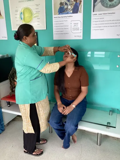
What is a fundus examination?
A fundus examination — you may also hear it called a fundus checkup or dilated fundus exam — is simply a careful look at the fundus, the back of your eye, where the retina, macula, optic nerve and blood vessels sit. Once a drop has widened the pupil, we use a bright light and a special lens to see right inside and pick up any problem early. It is the very same examination as our retina & macula check here at My Vision Eye Care.
A retina and macula examination is quick and comfortable, and it involves no intervention: just dilating drops and a careful look at the back of the eye. Depending on the drop used, the dilation may last four to five hours, or occasionally 24 to 48 hours.
At My Vision Eye Care we examine your eyes for any risk of disease developing. People with diabetes and hypertension are especially prone to retinopathy, and eyes that are short-sighted or already have macular disease carry their own risks. To help you stay ahead of any change, we give you a simple home-monitoring guide, so you can watch your own vision between visits and tell us the moment anything looks different.
Searching for a “fungus” eye check-up? You very likely mean a fundus examination — the words sound alike, but they are quite different things. A fundus exam is a routine look at the back of the eye, while a fungal eye infection is a separate and serious problem that needs quick care. We untangle the two in our guide: fundus examination vs “fungus” in the eye.
Retina & Macula — your questions answered
What is a fundus examination?
A fundus examination — also called a fundus checkup or dilated fundus exam — is a careful look at the fundus, the back of your eye where the retina, macula, optic nerve and blood vessels sit. Once a drop widens the pupil, we use a bright light and a special lens to see inside and catch any problem early. It is the same examination as our retina & macula check.
Is “fungus in the eye” the same as a fundus examination?
No — the words only sound alike. A fundus examination is a routine, painless look at the back of the eye. A fungal eye infection (“fungus in the eye”) is a separate and serious problem — usually after an eye injury or contact-lens misuse — that needs quick care. If your eye is red, painful or watering, please see an eye doctor straight away. We explain the difference in our fundus vs fungus guide.
How often should I have a fundus examination?
For most healthy adults, having it as part of a full eye check every one to two years is plenty. If you have diabetes, high blood pressure, strong short-sight, or an existing retinal or macular condition, you may need it yearly or more often — we’ll advise the right interval for your eyes.
What is macular disease?
The macula is a small part of the retina responsible for your central vision. Tasks such as threading a needle, reading small print, reading street signs and seeing fine detail all rely on a healthy macula.
What is macular degeneration?
Many older people develop age-related macular degeneration (AMD) as part of natural ageing — often without realising it until a vision problem appears or it’s found during an eye exam. There are two types: dry and wet. Wet (exudative/neovascular) AMD causes more severe vision loss; the earlier it is diagnosed and treated, the better the chance of preserving central vision.
What are the symptoms of macular degeneration?
- Dark areas or distortion in the central vision; if unchecked it can cause permanent central vision loss — but it never causes total blindness.
- It usually begins in one eye and may affect the other later.
- An eye examination identifies the risk and distinguishes dry from wet AMD.
Can I find out if I have a progressive macular problem?
Yes. After an eye examination, you’ll be taught how to use a simple test to self-monitor early macular degeneration at home.
Can macular degeneration be prevented?
Some eyes are predisposed, but these guidelines help prevent or slow AMD:
- Don’t smoke.
- Eat plenty of dark, leafy green vegetables such as spinach, plus fruit and nuts daily.
- Consider a multivitamin/multimineral supplement and fish or fish-oil.
- Exercise regularly and maintain a healthy weight.
- Keep blood pressure and cholesterol under control.
- Wear appropriate sunglasses outdoors to block UV and blue light.
- Have regular eye exams with an ophthalmologist.
What is retinal detachment?
Some eyes have weak areas in the retina that can cause it to pull away from the back of the eye — a retinal detachment. A detached retina does not function normally, and vision becomes blurry.
How is retinal detachment treated, and how would I know I have it?
Retinal detachment can only be treated with re-attachment surgery; untreated, it can cause irreversible blindness. It is diagnosed during an eye examination after full pupil dilation (sometimes with an ultrasound of the eye). Warning symptoms include seeing red dots, flashes of light, or a curtain-like shadow falling across the vision. Periodic dilated eye exams prevent it in most eyes.
Can retinal detachment be prevented?
In many cases, yes. Regular eye exams reveal early changes, and treating weak retinal areas helps prevent detachment. Use appropriate eye protection during risky activities — sports goggles with polycarbonate lenses, and protective eyewear when using machines, chemicals or tools, or while gardening. These special glasses are injury-resistant, unlike fragile ordinary plastic lenses.
What is diabetic and hypertensive retinopathy?
Consistently high blood sugar, or long-standing diabetes, starts to damage the eye — a condition called diabetic retinopathy that usually affects both eyes. Early on there are often no noticeable changes, but as it progresses it usually causes irreversible vision loss. Sharing your recent blood-sugar control history is important.
Can severe vision loss from diabetic retinopathy be prevented?
Yes. An eye examination is the only way to detect diabetic retinopathy, and periodic comprehensive exams monitor its progression. On examination, visual acuity is measured, the front of the eye is examined on the slit-lamp, and the dilated retina, macula and optic nerve are checked for damage — after which drops, laser or further tests may be advised.
What is the recommended diabetic eye-screening schedule?
The American Academy of Ophthalmology recommends:
- Type 1 diabetes: within five years of diagnosis, then yearly.
- Type 2 diabetes: at the time of diagnosis, then yearly.
- During pregnancy: pregnant women with diabetes should see their ophthalmologist in the first trimester, as retinopathy can progress quickly.
Related care
All services Low Vision Rehabilitation When vision loss can’t be reversed by glasses, medicine or surgery, rehabilitation helps you keep doing the everyday things that matter — independently.
  Eye Examination for All A thorough, all-ages eye screening that confirms the absence of any vision-threatening condition — and catches early disease before it steals your sight.